Here is the short answer first. T2 hyperintensity means a small area showed up brighter than the tissue around it on one particular MRI camera setting, called the T2 setting. On a brain scan these are sometimes described as "white spots." The brightness is a description of how the area looks in the picture. It is not a diagnosis, and on its own it does not tell you that anything is wrong.
T2 hyperintensities are extremely common, especially as we get older, and most of the time they are linked to ordinary, manageable things like the natural ageing of tiny blood vessels or a long history of migraines. Sometimes, depending on your age, how many spots there are, and whether they are new, your doctor will want to look a little closer. This guide explains what the term means, why these bright areas appear, the common reasons behind them, what the related word "FLAIR" means, why the location of the spots matters, and what your doctor actually weighs when reading them.
Now the part that probably brought you here. Your MRI report says "T2 hyperintensity" or "white matter hyperintensities," it reached your phone on WhatsApp before you could see the doctor, and your brain immediately jumped to the worst thing. "Spots on my brain." Take a breath. For a huge number of people, these are among the most common and most ordinary findings a radiologist reports.
Read this calmly, top to bottom. By the end you will know what the term describes, what it does not, and what to ask your doctor.
What does T2 hyperintensity mean on an MRI?
T2 hyperintensity refers to an area that appears brighter than the surrounding tissue on the T2 setting of an MRI scan. To unpack that, an MRI takes pictures using a few different camera settings, and T2 is one of them. On the T2 setting, anything with a lot of water or fluid in it tends to look bright. So a "T2 hyperintense" area is simply a spot that lit up brighter than its neighbours on that particular setting.
That is the whole literal meaning. It is a brightness description, not a verdict.
A T2 hyperintensity typically refers to a small area that holds slightly more fluid than the tissue around it and therefore looks brighter on the scan. The reason this matters is that "bright spot" sounds dramatic and usually is not. The brightness tells the radiologist where to look. It does not, by itself, tell anyone what the spot is. Your doctor will use this finding, alongside your full clinical picture, to determine what it means for you specifically.
Why do these bright areas show up on a brain MRI?
Bright areas show up on a T2 scan because of small differences in water content between one patch of tissue and the next. Wherever there is a little more fluid than usual, that patch glows brighter on the T2 setting. That is the entire mechanism. The MRI is not detecting "damage" as a concept, it is detecting water, and then the radiologist interprets what that pattern of water might represent.
In the brain, these bright patches most often appear in the white matter. White matter is the deeper part of the brain that acts like the wiring, the bundles of nerve fibres that carry signals between different areas. The outer surface layer is called grey matter. When a report mentions "white matter hyperintensities," it is describing bright spots in that deeper wiring layer.
Tiny shifts in fluid in the white matter are common, and they accumulate gently with age in almost everyone. So the appearance of some bright spots there is, very often, an expected part of a brain that has been used for a few decades. What the spots represent is the part your doctor interprets, not the scan itself.
What are the common causes of T2 hyperintensity?
Most T2 hyperintensities in the brain trace back to a small number of common, often manageable explanations, and only a minority point to something a doctor needs to chase further. The single most common explanation, especially from middle age onwards, is changes in the brain's tiny blood vessels, often written as "small vessel disease" or "chronic small vessel ischaemic changes." That phrase sounds frightening, but it usually describes gradual, age-related wear in the smallest blood vessels, and it is very widespread in people with high blood pressure or diabetes, both of which are common in India.
The second common association is migraine. People who have had migraines for years can show small white matter bright spots that are well recognised and generally not alarming on their own.
Beyond these, there is a wider set of less common explanations that doctors evaluate when the pattern, the patient's age, or the symptoms call for it. These can include inflammation, changes to the protective covering of nerves, or the after-effects of an old, healed event. None of these is something you can read off the report yourself.
Here is the safe way to hold all of this. T2 hyperintensity can be associated with several different things, ranging from ordinary age-related changes to causes that need follow-up. It is one of several pieces a doctor considers. It is not a diagnosis, and it does not tell you which cause applies to you. Your doctor decides that using your age, your history, your symptoms, and sometimes further tests.
What does FLAIR mean, and how is it different?
FLAIR is a closely related MRI setting, and you will often see the phrase "T2 FLAIR hyperintensity" in a report. FLAIR stands for Fluid Attenuated Inversion Recovery. The long name describes one clever trick: it is a T2-style setting that deliberately dims down the normal fluid in the brain, like the fluid in the central spaces, so that genuine spots stand out more clearly instead of being lost against bright background fluid.
So "T2 FLAIR hyperintensity" typically refers to a bright area seen on that fluid-dimmed setting, where it is easier to spot. It is a description of how the area appears under a specific setting, not a separate or scarier finding.
In practice, radiologists rely on FLAIR a lot for the brain because it makes white matter spots easier to see near the fluid-filled spaces. If your report mentions both T2 and FLAIR, it usually just means the radiologist confirmed the same bright area on more than one setting. That is normal and thorough, not a sign of something worse. As always, what the area represents is your doctor's call.
Why does the location of the spots matter?
The location of a T2 hyperintensity matters because the same brightness can mean different things depending on where in the brain it sits, which is why radiologists describe the position so precisely. You will see location words like periventricular, deep white matter, and cortical, and they are simpler than they sound.
| Location word | What it means in plain language |
|---|---|
| Periventricular | Near the ventricles, the fluid-filled spaces in the centre of the brain |
| Deep white matter | Deeper inside the wiring layer, away from the surface |
| Cortical | At or near the brain's outer surface layer |
The radiologist notes location because the pattern helps your doctor narrow down the likely explanation. Spots in one distribution can point gently towards ordinary age and blood-vessel changes, while a different distribution might prompt a doctor to consider other causes and look closer.
The important thing for you as a patient is this: the location is information for your doctor, not a code you are meant to crack at home. A location word in your report is the radiologist being precise, so your doctor can interpret the pattern properly. It is not a hidden severity rating.
How worried should I be? What your doctor actually weighs
The honest answer is that a T2 hyperintensity on its own does not come with a built-in worry level, which is exactly why your doctor, not the report and not the internet, is the one who reads it for you. What looks identical on a scan can be completely ordinary in one person and worth following up in another, because the meaning depends on the person, not just the picture.
Here is what a doctor genuinely weighs, so you understand the thinking rather than guessing at it:
- Your age. Some bright spots are an expected part of an older brain and unremarkable.
- One spot versus many. A single small spot and a scattered pattern of many spots are read differently.
- New versus old. If you have had previous scans, your doctor compares whether anything has changed.
- Your symptoms. The same finding is interpreted differently in someone with no symptoms than in someone with specific complaints.
- Your history. Blood pressure, blood sugar, migraines, and past events all shape the reading.
Notice what this means. A small, single, stable spot in an older adult with high blood pressure is a very different conversation from several new spots in a young person with new symptoms, even though both reports use the words "T2 hyperintensity." You cannot place yourself on that spectrum from the report alone, and you should not try to. That placement is your doctor's job, and they do it with the whole picture in front of them.
What should you do after reading this on your report?
After seeing "T2 hyperintensity" on your report, the most useful thing you can do is resist the urge to search that one phrase in isolation, read your whole report calmly, and take clear questions to your doctor. A single line out of a full report is not your situation, and read alone it almost always sounds worse than it is.
Calm questions to carry to your doctor:
- In plain words, what do you think this T2 hyperintensity represents in my case?
- Given my age and history, is this something we act on or simply note?
- Do my symptoms, if any, connect to this finding?
- Do I need a follow-up scan to compare over time?
- Is there anything reassuring here that I might have missed?
Bring the full report, not a screenshot of one line. Give your doctor the whole picture.
How FlexReport helps you understand your report
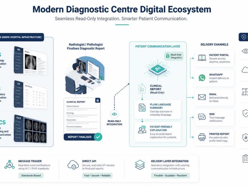
FlexReport is a patient comprehension layer for diagnostic centres, labs, and hospitals. It adds a plain-language second view to clinical reports without altering the clinical report itself. FlexReport operates in India and is built by FlexRad, a unified radiology platform with imaging infrastructure across India and the US.
In plain terms: your radiologist's report stays exactly as written, because that is the official record. FlexReport sits alongside it and adds a simplified explanation, often on the same WhatsApp thread or patient app your diagnostic centre already uses. A phrase like "T2 FLAIR hyperintensity in the periventricular white matter" stops being a wall of jargon and becomes something you can actually read.
What FlexReport does not do is diagnose you, replace your doctor, or tell you what your treatment should be. That stays with your treating physician, where it belongs.
For the full walkthrough of every part of an MRI report, see our guide on how to read an MRI report.