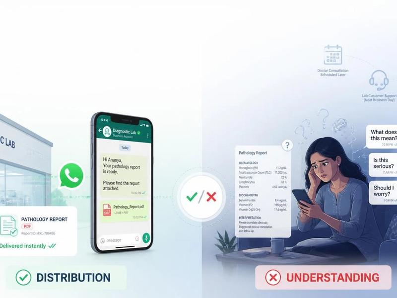
You got your scan report on WhatsApp. You opened it before the doctor could call. And now you are reading words like heterogeneous, lesion and degenerative changes, and your stomach has dropped.
Take a breath. This page exists for exactly this moment. It is a plain-language glossary of the terms that show up on an MRI (Magnetic Resonance Imaging), CT (Computed Tomography), X-ray and ultrasound report. No medical degree needed. Most of these words are simply describing what the scan picture looks like. They are not telling you what is wrong with you. That part is your doctor's job, and you will get to it soon.
Find your word below, read the one or two lines, and breathe a little easier until you see your doctor. If you would rather not hunt term by term, FlexReport can take your whole report and give you a plain-language version of it in a few minutes.
Read this first: your report describes, it does not decide
Here is the single most useful thing to understand before you read a single term. A radiology report is a description of a photograph. The radiologist is writing down what the image looks like, the same way you might describe a photo of a room: this corner is darker, that shape is round, this edge is blurry. None of those descriptions, on their own, are a diagnosis.
So every word in this glossary works the same way. The term typically refers to a feature on the scan. It is a description, not a diagnosis. Your treating doctor reads this report alongside your symptoms, your history, your blood tests and a physical check, and only then decides what it actually means for you.
A scary-sounding word in isolation is just a word in isolation. Keep that frame as you read, and most of the fear drains out of these reports.
How a radiology report is structured, and which part actually matters
Before the words, understand the shape. Almost every radiology report follows the same five parts, in the same order, no matter which centre issued it.
| Section | What it refers to |
|---|---|
| Clinical History (or Indication) | Why the scan was done in the first place. |
| Technique | How it was done: which machine, and whether dye was used. |
| Comparison | Whether your scan was checked against an older one. |
| Findings | The long, detailed middle section listing everything noticed, including all the normal things. This is where the frightening words live, and it looks alarming precisely because it records everything. |
| Impression | The short summary at the very end. The part your doctor reads first and cares about most, because it is the radiologist's bottom line. |
What this glossary really gives you
This glossary is a plain-language dictionary of the words on scan reports. Some people search for it as an imaging findings glossary or a scan report glossary. Whatever you call it, the job is the same: take a technical word like hypoechoic or focal or disc bulge, and tell you in one or two everyday lines what it is describing on the image.
You need this because of timing. In most cases you receive the report before you sit in front of the doctor who ordered it. That gap, sometimes a few hours, sometimes a day, is when the worry happens. You read a word you do not know, you type it into Google, and the internet hands you the single scariest possible meaning. A glossary built for patients does the opposite. It gives you the calm, ordinary meaning first, so you can wait for your appointment without spiralling. Think of this as the friend who happens to know hospital words, sitting next to you while you read.
The words, grouped so you can find yours fast
These are grouped the way a worried person actually thinks, not the way a textbook would. Find the group that matches your word.
Words that describe how something looks
These are texture and brightness words. They describe the appearance of an area, nothing more.
| Term | What it describes |
|---|---|
| Heterogeneous | An area that does not look the same all over; a mixed or patchy appearance. |
| Homogeneous | An area that looks uniform, the same throughout. |
| Hypoechoic / Hyperechoic (ultrasound) | Hypoechoic shows up darker on the scan; hyperechoic is the brighter version. "Echo" just means the sound wave bouncing back. |
| Hypodense / Hyperdense (CT) | Hypodense looks darker or less solid on a CT; hyperdense is the brighter, denser version. |
| Hypointense / Hyperintense (MRI) | Hypointense looks darker on an MRI; hyperintense is the brighter version. |
| Ill-defined / Well-defined | Ill-defined edges are blurry or unclear; well-defined edges are clean and sharp. |
Words for a spot, a lump or an area
These are the words that scare people the most and usually need to the least.
| Term | What it describes |
|---|---|
| Lesion | Any area that looks different from the tissue around it. That is the whole meaning. A pimple is technically a skin lesion. |
| Nodule | A small rounded spot, usually under three centimetres. |
| Mass | A larger area, usually over three centimetres. It is a size word, not a verdict. |
| Cyst | A sac filled with fluid. Extremely common, and most are harmless. |
| Focal / Diffuse | Focal is something in one specific spot; diffuse is something spread across an area. |
| Incidental finding | Something the scan happened to pick up that you were not even being checked for. Often minor, noted simply so it stays on record. |
Words you will see on back and spine reports
If your report is for back or neck pain, this is your section. These words appear on a huge share of Indian spine reports, and most describe normal ageing.
| Term | What it describes |
|---|---|
| Disc bulge | The soft cushion between two back bones quietly pushing outward a little. Very common after thirty, often in people with no pain at all. |
| Disc herniation (prolapse, slipped disc) | That cushion pushing out further, sometimes near a nerve. |
| Degenerative changes | Normal age-related wear and tear of the spine. |
| Spondylosis | Simply the medical name for that age-related spine wear. |
| L5-S1, L4-L5 | The names of the joints in your lower back. L stands for lumbar (the lower back bones), S for sacrum (the bone at the base of the spine). So L5-S1 is just an address for one joint. |
| Annular tear | A small crack in the tough outer ring of a spinal disc. |
| Canal stenosis / foraminal narrowing | The channel that nerves pass through becoming a little narrower. |
Direction and position words
These tell the doctor where on the body something is. They are map words, with no good or bad meaning attached.
| Term | What it describes |
|---|---|
| Anterior / Posterior | Front / back. |
| Medial / Lateral | Medial is the side closer to the middle of the body; lateral is the side toward the outer edge. |
| Proximal / Distal | Proximal is the part closer to where a limb joins the body; distal is farther away, toward the hand or foot. |
| Superior / Inferior | Higher up / lower down. Nothing to do with quality, only position. |
MRI image-setting words
These look like findings but are mostly just names for the camera settings the MRI uses.
| Term | What it describes |
|---|---|
| T1 and T2 | Two different settings the MRI machine uses to take pictures. The same body part looks different on each, which is why you often see both. They are settings, not findings. So "T2 hyperintense" simply means an area looked brighter on the T2 setting. |
| FLAIR (Fluid-Attenuated Inversion Recovery) | A special MRI setting that dims normal fluid so other things stand out. Again, a setting name. |
| Enhancement | An area lighting up more after the contrast dye is given. |
| Attenuation | How much the scan beam is absorbed, which decides how bright or dark an area appears on a CT. |
Words for fluid, swelling and inflammation
| Term | What it describes |
|---|---|
| Edema | Swelling caused by extra fluid collecting in tissue. |
| Effusion | A pocket of fluid gathering in a space, often around a joint or a lung. |
| Inflammation | The body's normal response to irritation or injury. |
| Opacity (chest X-ray) | An area of lung that looks whiter or cloudier than the rest. Many possible causes. |
| Consolidation | A part of the lung that has filled with fluid or other material instead of air. |
Other findings words you might see
| Term | What it describes |
|---|---|
| Sclerosis | An area, often bone, that has become harder or denser than usual. |
| Stenosis | A narrowing of a passage, tube or channel. |
| Atrophy | A part that has shrunk or thinned, often with age or reduced use. |
| Calcification | Tiny calcium deposits, like small specks of bone-like material. Very common and often harmless. |
| Mass effect | One structure gently pushing on or shifting another nearby. |
| Necrosis | An area of tissue that is no longer active. This is one your doctor will always interpret in full context, so do not read it alone. |
Words that sound scary but are usually calm news
| Term | What it describes |
|---|---|
| Unremarkable | Nothing abnormal being seen. Genuinely the best word in any report. |
| No acute abnormality | Nothing urgent or new being found. |
| Benign | Something that is not cancer and not harmful. |
| Prominent | Something slightly larger than average, which is often just how you are built. |
| Artefact | A false mark on the image caused by movement or the machine, not something inside your body. |
| Within normal limits | Simply normal. |
Words about the dye and the scan itself
| Term | What it describes |
|---|---|
| Contrast (dye) | A safe liquid given to you so certain areas show up more clearly. |
| With and without contrast | A scan done twice, once before the dye and once after, so the two can be compared. |
| Plain study | A scan done without any dye. |
The Indian reality: you can see a doctor fast, and you still sit with the fear
Here is the part most foreign health websites get wrong about us. They tell you to wait calmly for your follow-up appointment in five days. In India, that is not how it works. If you are anxious about a report, you do not wait. You walk into the neighbourhood clinic, you ask a known doctor, or you simply go to a second doctor two lanes away within the hour. Access to a doctor is rarely the problem here.
The fear in the waiting is still the problem. Because even if your appointment is only a couple of hours away, those couple of hours are spent staring at words you cannot read. The report is already on the family WhatsApp group. An uncle has Googled one term and announced his theory. Your mother is quietly worried in the next room.
By the time you reach the doctor, the whole family has lived through a crisis that the report never actually described.
That is the gap this page is for. Not to replace your doctor, who you will see soon, but to keep you steady in the hours before you do. Read your terms here, understand that they are descriptions, and walk into that clinic with questions instead of panic.
Questions to ask your doctor after your scan
Walking in with questions changes the whole appointment. Here are six worth writing down before you go. They turn a one-line answer into a conversation you can actually use.
- What does the Impression at the end of my report mean in plain words?
- Is anything in this report something to act on now, or something to simply watch over time?
- Are the findings here normal for my age, or unusual?
- Do I need another test or scan to be sure, and if so, why?
- If there is a finding, what are the possible explanations, and which is most likely in my case?
- What would make you want to see me again sooner rather than later?
Save this list, or screenshot it before your visit. A doctor would far rather answer six clear questions than calm down a patient who has spent the morning reading the worst corner of the internet.
Do I still need to see my doctor after reading this?
Yes, completely. This glossary helps you understand the words in your report. It does not tell you what your report means for your body, your treatment or your next step. Only your treating doctor can do that, because only your doctor has the rest of the picture: your symptoms, your history, your examination and your other tests.
Think of it like this. This page helps you read the map. Your doctor is the one who knows the destination and the road. Understanding the words ahead of time does not shorten the journey, but it does mean you arrive at the doctor's room able to ask better questions and follow the answers. That is the entire goal: calmer patient, clearer conversation, better appointment.
How to get your whole report in plain language
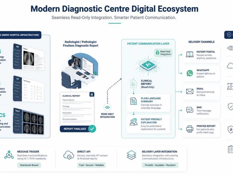
FlexReport is a patient comprehension layer used by diagnostic centres, labs and hospitals. It adds a plain-language second view to the report you already received, without changing a single word of the original clinical report your centre issued. It is built in India by FlexRad, a unified radiology platform that handles the storage and sharing of scan images across India and the US.
In plain terms: instead of you decoding your report one word at a time, FlexReport's Engine reads the whole thing and gives you a simplified version you can actually follow, on the same WhatsApp thread your centre already uses.